


Nail psoriasis is a special form of psoriasis in which the fingernails and / or toenails are affected. Doctors call this type of disease psoriatic onychodystrophy (from the Greek.onychos- clove,dis- rape,tropic- food).
From this article, you will learn about the causes of the development of nail psoriasis, its symptoms, which do not always unequivocally indicate the correct diagnosis, as well as dangerous misconceptions about this form of the disease.
Note.There are many photographs in the article that may scare an unsuspecting reader.
Where do nails grow from?
To understand the problem of nail psoriasis, it is important to understand how the so-called nail apparatus works.
The nail has two functions: functional and aesthetic. First of all, the nail protects the fingertips from damage, increases precision and sensitivity when working with small objects, it can be a weapon of attack or defense, and finally, with the help of nails we bite. Second, the aesthetic or cosmetic function of the nails is also important, especially for women.
Nails are formed from the outer layer of the skin, the epidermis. The nail apparatus includes:
- nail plate: directly the nail itself,
- matrix: produces the nail plate,
- the nail hole, or lunula, is the only visible part of the matrix, this is a white moon-shaped area at the base of the nail plate,
- eponychium - a nail roller that protects the matrix from above from damage,
- nail bed - located under the nail plate and is responsible for its attachment to the nail phalanx of the finger,
- hyponychium: the transition zone between the nail bed and the skin of the fingertip.
Causes and mechanism of development of nail psoriasis.
In its course, with periodic exacerbations and remissions, nail psoriasis resembles a vulgar form of the disease.
Nail psoriasis is believed to develop for the same reasons and in the same pattern as typical psoriatic rashes. Among these reasons, external and internal factors are distinguished.
The main intrinsic factor is genetic predisposition. External causes are numerous and include, for example, injuries, poor diet, intoxicants (alcohol and tobacco), infections, and certain medications.
The standard mechanism of development of nail psoriasis under the influence of these reasons can be briefly described as follows:
- Triggering factors, such as trauma, activate immune cells.
- Activated immune cells migrate to the nail matrix area or the nail bed.
- Immune inflammation develops in these areas.
- The division of skin cells is abruptly accelerated and their maturation is interrupted.
- There are characteristic symptoms of nail psoriasis.
Also, the cause of nail psoriasis can be considered as a result of the body's inability to adapt to adverse environmental conditions. According to this view, the main cause of psoriasis is an evolutionarily strange habitat.
As a consequence, this evolutionary approach considers unhealthy eating, lack of sunshine and clean water, excess toxins, lack of normal physical activity, sleep disturbances, and chronic stress as direct causes of the disease.
Nail psoriasis and psoriatic arthritis are related
The connection between nail damage and psoriatic arthritis has been known for a long time.
Based on observations, scientists have found that psoriatic arthritis is accompanied by nail damage in nine out of ten cases.
But the mechanism of this connection has not been fully studied. However, the authors of several studies, for example from the Leeds Institute of Molecular Medicine (UK), tried to explain this connection beyond the concept of immune inflammation.
In his opinion, the truth is that the finger joint is located next to the nail and is anatomically associated with it.
Therefore, microtrauma and Kebner phenomenon that cause primary joint inflammation (psoriatic arthritis) also cause secondary pathological changes in the nearest nail.
This is why psoriatic arthritis is associated with nail damage so often.
%20of%20the%20toes.jpg)
Therefore, the symptoms of nail psoriasis are often indicative of psoriatic arthritis.
Let's now look at the main myths that accompany this disease and how dangerous they are.
Myth 1: nail psoriasis is rare.
Not really. Apparently, with psoriasis, the nails suffer very frequently.
According to various sources, nail psoriasis occurs in the range of 6% to 82% of cases of psoriasis vulgaris. Such a wide extension in the evaluation of the prevalence of this pathology is explained by problems in its accounting. Medical statistics record visits to doctors firstly by patients with a vulgar form, and secondly, attention is paid to nails. In scientific research, nail psoriasis cases are also generally studied only in addition to the main object of interest - psoriasis with skin lesions.
However, several publications say that
up to 80-90% of patients with psoriasis vulgaris reported recurrent nail damage.
And also that nail psoriasis occurs in 90% of patients with psoriatic arthritis and scalp psoriasis.
It should be noted that, generally, adults suffer from this form of the disease.
According to various sources, in children, nails are affected in approximately 7-37% of psoriasis cases. Unfortunately, the manifestations of psoriasis on the nails of a child are often not given due importance. Parents or doctors believe that this is a variant of the norm or a consequence of trauma, or they simply don't realize it due to the mild severity of the symptoms.
Myth 2: recognizing nail psoriasis by symptoms is easy
In fact, not always. The fact is that
the nail can respond to various diseases with only a limited number of symptoms. Therefore, the manifestations of various nail diseases can look the same.
Of course, nail psoriasis can be suspected if the patient has severe symptoms of psoriasis vulgaris. However, nail injuries can be minor compared to skin injuries and can be easily ignored by a doctor.
Generally, the more active psoriasis is on the skin, the more severe the damage to the nails.
First of all, the nails are affected.
And it is also important to know that in 5% of cases, the nails can be the only initial manifestation of psoriasis. That is, the classic manifestations of psoriasis on the skin may be completely absent.
The appearance of nail psoriasis depends on where the pathological changes originate: in the matrix or in the nail bed.
It is important to consider the source of symptoms (womb or bed) when choosing a treatment. Therefore, it is necessary to define it correctly.
The symptoms that originate in the nail matrix are:
- thimble symptom,
- white spots and spots (leukonychia),
- red dots in the hole,
- crumbling nails.
Although the cause of these symptoms is at the level of the matrix, as the nail grows, pathological changes appear in the nail plate.
The symptoms, the cause of which is in the nail bed, are:
- detachment of the nail (onycholysis),
- longitudinal bleeding
- subungual hyperkeratosis,
- symptom of an oil stain.
Next, we will look at each symptom separately. And let's start with the manifestations that originate from the womb.
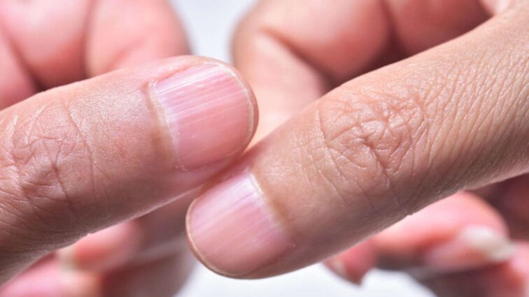
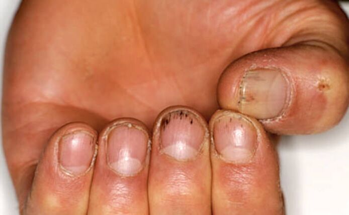
Thimble symptom
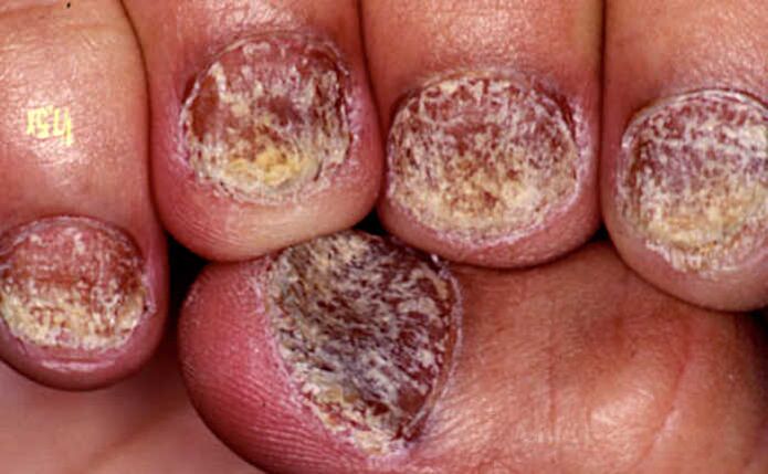
The symptom of a thimble appears on the surface of the nail plate with holes or pits, which look like depressions in a thimble.
Such defects mainly occur on the fingernails, but rarely appear on the feet. As the nail grows, the holes move from the nail fold to the edge of the nail plate.
The pits in nail psoriasis are usually deep, large, and chaotic. They arise due to the detachment of loose groups of cells from the surface of the nail, in which division and keratinization are altered.
The more severe the psoriasis, the more often the thimble symptom occurs.
However, it must be borne in mind that, in addition to psoriasis, nail pits are also characteristic of alopecia areata (alopecia), eczema, dermatitis, and can also occur, for example, with a fungal infection.
Counting the total number of holes in all nails will help to make a correct diagnosis.
- Less than 20: not typical for psoriasis,
- 20 to 60 - psoriasis may be suspected,
- Over 60 - Confirm the diagnosis of psoriasis.
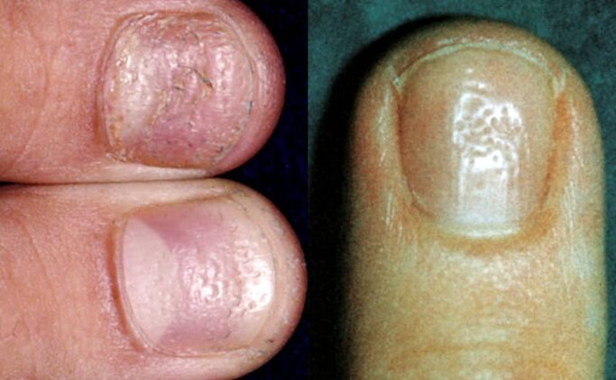
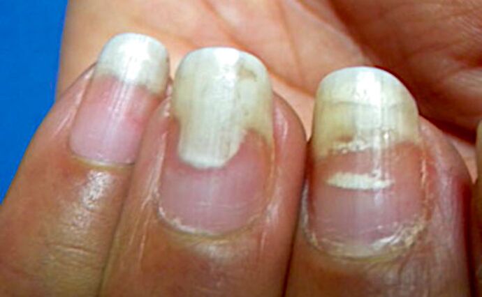
White spots (leukonychia)
Leukonychia is a symptom that manifests itself as white spots or dots on the nails.
With leukonychia (from the Greek.leukós- White andonychos- nail), in contrast to the superficial pits in the symptom of a thimble, cells with impaired division and keratinization are found in the thickness of the nail plate. At the same time, the nail surface remains smooth. And the white color of the spots arises from the reflection of the light of the groups of cells little located.
However, some studies suggest that leukonychia is so common in healthy people that it is not a characteristic symptom of psoriasis. For example, a manicure injury can cause leukonychia.
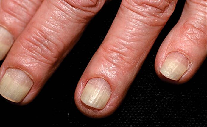
Crumbled nails
When the superficial pits (thimble symptom) and deep areas of leukonychia (white spots) merge, the nails begin to crumble.
Nail shedding usually occurs with prolonged nail psoriasis.
And the more intense the inflammation of the nail matrix, the more the nail plate is destroyed. In severe cases, the nail can completely collapse and fall off.
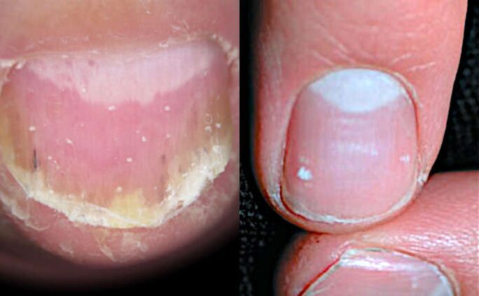
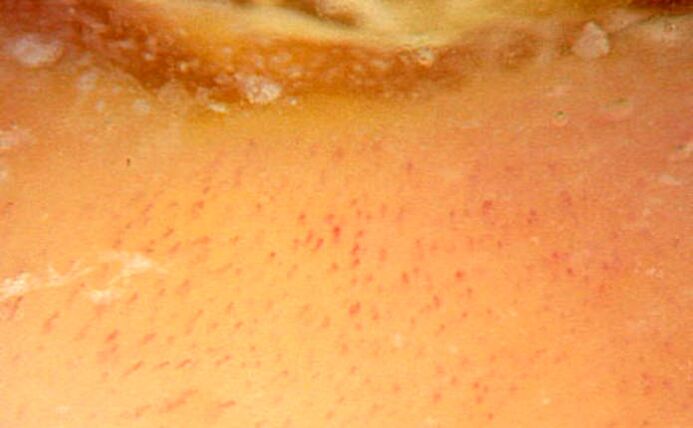
Red spots in the nail socket.
Apparently, the red dots in the area of the hole and its general redness occur due to increased blood flow to the vessels under the nail.
Also, red dots are formed in the hole due to a violation of the structure of the nail plate - it becomes more transparent and thinner. And because of this, firstly, the vessels become more visible, and secondly, the thin nail plate puts less pressure on the vessels under it and they are more filled with blood.
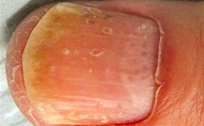
Nail plate thinning can also cause redness of the entire nail bed.
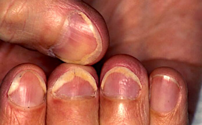
Nail detachment (onycholysis)
Now let's consider the symptoms, the source of which is the nail bed.
Onycholysis is the separation of the nail plate from the bed due to the accumulation of cells under the nail with impaired division and keratinization.
Onycholysis itself (from the Greek.onychos- nail andλύσις- separation) is not necessarily a sign of psoriasis and can develop, for example, as a result of a nail injury.
Initially, the loss of contact between the nail and the bed occurs in the area of hyponychia, along the outer edge of the nail plate. Then the onycholysis extends into the nail fold in a semicircular line. The peel area turns white due to the accumulation of air under the nail.
A reddish border (scientifically erythema) along the edge of the onycholysis, which is usually visible on the fingers, is characteristic of psoriasis and helps to make a correct diagnosis.
With prolonged onycholysis, the nail bed loses its properties and the newly growing nail will most likely not be able to adhere to it normally. Therefore, even with a complete renewal of the nail plate, onycholysis often persists.
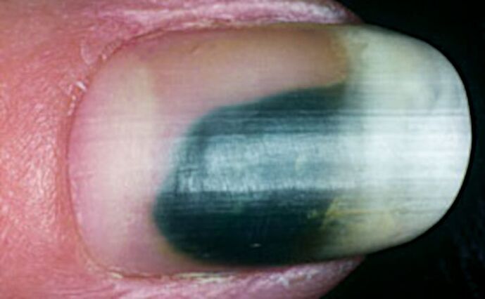
Due to the fact that onycholysis facilitates the penetration of bacteria and fungi, the infection can join. This sometimes leads to discoloration of the nail. For example, a greenish color may appear when bacteria adherePseudomonas aeruginosa(Pseudomonas aeruginosa) and others.
Longitudinal subungual hemorrhage
Longitudinal subungual hemorrhages occur in the nail bed and appear as dark red lines 1-3 mm long.
Increased blood flow and edema in the area of inflammation of the nail bed lead to the rupture of capillaries, which manifests itself in the form of such hemorrhages.
Due to the peculiarities of the blood supply, most hemorrhages occur closer to the free edge of the nail, in the area of hyponychia.
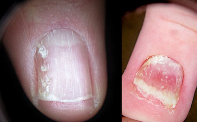
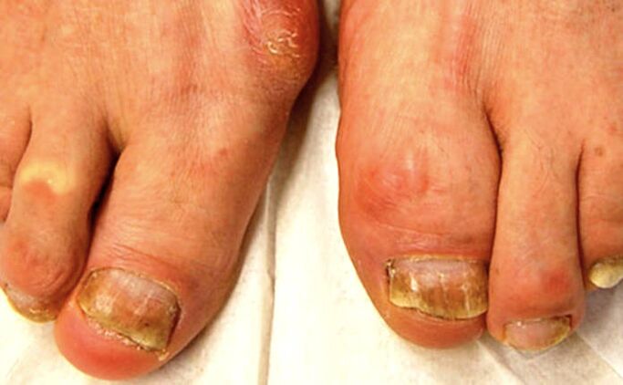
Subungual hyperkeratosis
Subungual hyperkeratosis is a collection of dead cells under the outside of the nail plate.
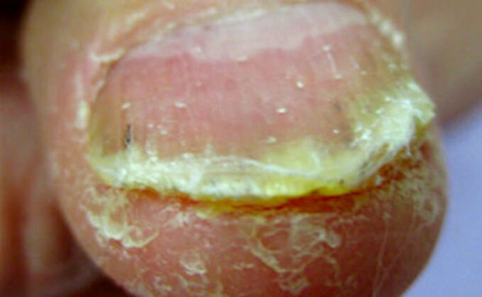
With psoriasis, subungual hyperkeratosis (from the Greek.hyper- excessively andkeras- horn) is usually silvery-white in color, but can also be yellow. And when the infection adds up, it can turn, for example, greenish or brown.
The higher the nail is raised above the nail bed, the greater the activity of the pathological process.
In the fingers, subungual hyperkeratosis usually manifests as loose layers under the nail plate. On the legs, these masses are strongly welded with a thickened nail.
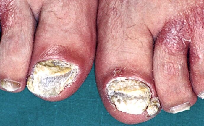
In addition, psoriasis with toenail lesions is characterized by a combination of subungual hyperkeratosis with onycholysis (separation of the nail).
Oil stain symptom
The symptom of an oil stain appears under the nail plate in yellow-red (salmon) patches.
They arise in the nail bed closer to the nail fold and move towards the edge of the nail as it grows.
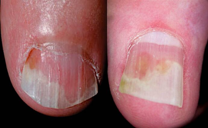
The cause of this symptom is inflammation of the nail bed with expansion of capillaries and accumulation of cells involved in inflammation, as well as cells with impaired division and keratinization.
Oil stains come in a variety of shapes and sizes. They can be found both in the center of the nail and on the edge, next to the onycholysis area.
Myth 3: nail psoriasis is just a cosmetic problem.
In fact, this is not true. Although more than 90% of patients report unsightly psoriasis nails, this is not just a cosmetic issue.
According to various studies, nail psoriasis significantly reduces the quality of life of patients:
- 52% of patients also complain of pain,
- 59% - for problems in daily activities,
- 56% - due to difficulties at home and
- 48% - for difficulties at work.
Therefore, it is very important to make the correct diagnosis and start treatment as soon as possible, since improving the condition of the nails significantly improves the quality of life of patients with psoriasis.
Myth 4: nail psoriasis is not dangerous
Actually, this is not the case. Speaking earlier about the causes of this form of the disease, we have already written that
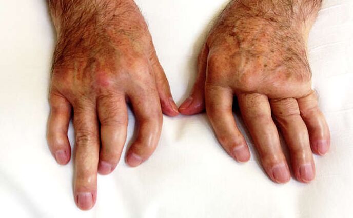
Nail psoriasis is a major symptom of psoriatic arthritis.
It is important to note that the external manifestations of arthritis may be completely absent. In this case, we can talk about not only the fact that the joints of the fingers and toes are affected, but also the joints of the spine and pelvic bones may be affected.
You can check your joints for arthritis with ultrasound (ultrasound) or magnetic resonance imaging (MRI).
Even if there are no obvious symptoms of arthritis, but there are manifestations of nail psoriasis, it is very important to make sure that all the joints are in order.
And then regularly check the condition of the joints. Otherwise, psoriatic arthritis can easily be overlooked! A late diagnosis will lead to late treatment and, as a result, irreversible joint damage and disability.
Therefore, if the doctor has not ordered an insurance investigation, citing the absence of visible signs of arthritis, you should contact the clinic yourself and undergo, for example, a paid ultrasound.
How to diagnose nail psoriasis
It is important to be able to recognize the many symptoms of nail psoriasis, which we described above, as they help to establish the correct diagnosis. But since the characteristic nail changes of psoriasis can also occur in other diseases, it can be difficult to make a correct diagnosis right away.
In this case, the presence of several symptoms at the same time in different nails can help in the diagnosis.
The important signs of nail psoriasis are:
- a symptom of a thimble: more than 20 holes in all fingernails indicate the possibility of psoriasis, and more than 60 holes confirm the diagnosis of psoriasis,
- detachment of the nail (onycholysis) with a reddish border around the edge,
- oil stains (salmon) on the nail bed.
Difficulties in diagnosing nail psoriasis from a single symptom
It is especially difficult to diagnose nail psoriasis if it presents with only one symptom.
For example, if there is only onycholysis in the hands or only subungual hyperkeratosis in the arms and / or legs.
The only method to make a reliable diagnosis in isolated onycholysis (nail detachment) is probably the study of hyponychia using a special microscope - a dermatoscope.
To do this, a high-magnification videodermatoscope is used. Note that the handheld dermatoscope does not provide the required magnification. What is needed is a videodermatoscope with a magnification of at least 40 times. The dilated capillary loops characteristic of psoriasis then become visible.
With isolated subungual hyperkeratosis, the likelihood of psoriasis is high if the accumulation of scales under the nail is silvery-white in color, as well as if all the fingernails or toenails are affected.
Psoriasis or nail fungus?
About 30% of nail psoriasis patients also have a fungal infection, scientifically onychomycosis.
Externally, hyperkeratosis and onycholysis (separation of the nail) in psoriasis can resemble the manifestations of a fungal infection. Therefore, it can be difficult to perform differential diagnoses, that is, to identify the true cause of the changes in the nail plate.
Also, both psoriasis and fungus can affect the same nails at the same time. Most of the time it occurs on the toes and is mainly characteristic of older patients.
Also, with a fungal infection, one or both of the big toe nails are often affected. In psoriasis, as a rule, several nails are affected at the same time.
The following symptoms speak in favor of psoriasis:
- oil stains and / or symptoms of a thimble on the nails,
- signs of psoriasis on the scalp and / or large skin folds,
- periodic remission and exacerbation of nail damage.
In favor of onychomycosis they say:
- longitudinal stripes on the affected nail,
- detection of fungi during microscopic examination of a scraping treated with potassium hydroxide from an affected nail (KOH test),
- positive culture for fungi.
In general, based only on external manifestations, it is impossible to completely exclude fungal nail infection in patients with psoriasis.
It should also be remembered that a fungal infection can cause Kebner phenomenon on the nail and the surrounding skin, resulting in psoriasis symptoms. Either way
It is helpful to go to a mycologist and do a test for fungi and, if found, start antifungal therapy.
Important findings and what to do
Let's summarize important information about nail psoriasis and its symptoms.
Diagnostic Features:
- Nail psoriasis is very common, but is often overlooked.
- Manifestations of nail psoriasis can be minor, so even experts often do not pay attention to it.
- In 5% of cases, nail damage may be the only symptom of early psoriasis.
- The manifestations of different nail diseases can look the same, which further complicates the diagnosis.
The main manifestations of nail psoriasis:
- a symptom of a thimble: holes in the nail,
- white dots,
- crumbled nails
- red dots in the hole area,
- detachment of the nail,
- longitudinal subungual hemorrhage,
- subungual hyperkeratosis: loose clusters under the nail,
- symptom of an oil stain.
Psoriasis and fungi:
- Nail psoriasis is often accompanied by a fungal infection.
- To unequivocally exclude it, it is necessary to contact a mycologist and conduct further research.
Nail psoriasis and psoriatic arthritis:
- Nail psoriasis is a common companion of psoriatic arthritis.
- It is important to detect pathological changes in the joints as soon as possible to start treatment in time and avoid irreversible complications and disability.
- Even if there are no external symptoms of arthritis, but nail psoriasis is detected, it is necessary to undergo an examination of the joints by ultrasound or MRI.